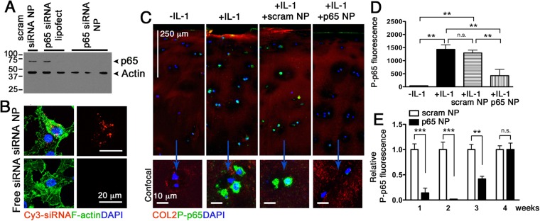
Figure 1.
p5RHH-p65 siRNA NP transfects HUVECs, human chondrocytes, and cartilage explants to suppress p65. (A) HUVECs were cultured to 50% confluence and transfected with p65 siRNA (RELA SMART pool, 100 μM F.C.) or scrambled siRNA delivered byLipofectamine or peptide-siRNA nanocomplex. p65 expression was detected by Western blot. Actin served as protein loading control. (B) Human primary chondrocytes were cultured as detailed in Materials and Methods and transfected with p5RHH-Cy3–labeled siRNA NP or free Cy3-labeled siRNA for 48 h. Cy3-siRNA (red) in chondrocytes was visualized by confocal microscopy. IL-1β stimulated human cartilage explants were incubated with p5RHH-p65 siRNA (p65 NP with 500 nM siRNA) or scrambled siRNA (scram NP) for 48 h after which the excess NP was washed off and cartilage stained and analyzed or cultured in the presence of IL-1β (10 ng/ml) for a total of 4 weeks. (C) Immunofluorescence of p65 (green) in cartilage sections at day 7 following different treatments. Bottom row: representative 2D confocal images at higher magnification. (D) Mean fluorescence of P-p65 per chondrocyte (from z-stack confocal images) 48 h after IL-1 β and NP exposure. (E) Relative mean fluorescence of P-p65 per chondrocyte at different time points. Mean P-p65 fluorescence in cartilage exposed to scrambled siRNA NP was set at 1. Mean reflects analysis of 5–8 areas per explant, each including 3–6 cells; 3–5 individual cartilage explants were analyzed per treatment per time point. **P < 0.01, ***P < 0.001; n.s. not significant. COL2 (red), type II collagen; DAPI (blue) stained nuclei.

