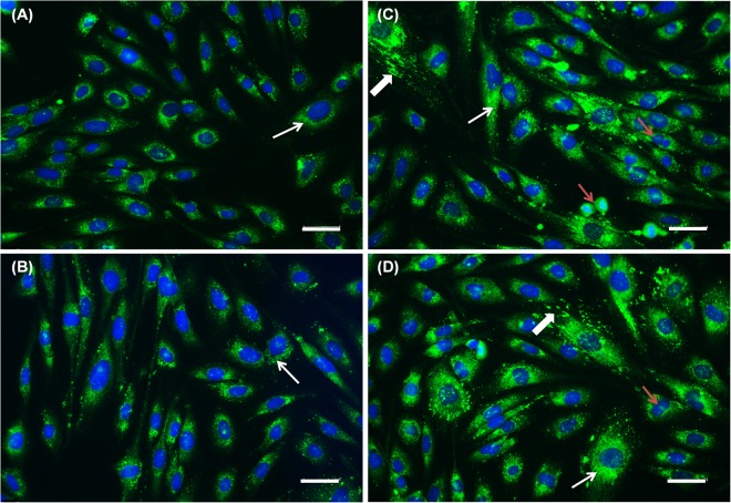
Figure 7.
Immunolocalization of type I collagen in fibroblasts treated for 48 h with (A) control (cell culture medium), (B) 100 μM AA solution, (C,D) DRV5 containing 100 μM AA. Thin arrows evidence positive intracellular labeling for type I collagen. Thick arrows evidence intense labeling of collagen I in the extracellular matrix. Red arrows show multiple dividing cells in the groups treated with (B) AA solution and (C,D) DRV5. Collagen was labeled with Alexa Fluor® 488 and nucleus with DAPI. Cells were analyzed by fluorescence microscopy with a 20x objective. Scale bars = 50 μm.

