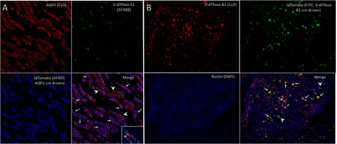
Figure 4.
(A) Triple immunofluorescence labeling of a kidney section from AQP2-cre+tdTomato+ (PC reporter) mice showing: AQP2 (red cells, upper left);. V-ATPase E1 (green cells, ICs, upper right); tdTomato (blue cells, lower left) staining in inner medulla. On the merged image (lower right) most AQP2 cells were also positive for tdTomato (purple cells, white arrows) while some were negative for tdTomato (red cells, white arrowheads). The dashed box contains a tubule imaged from another kidney section that contained an isolated cell that was positive for both AQP2 and V-ATPAse E1 (Yellow cell, asterisk). Magnification 40X, confocal microscopy. (B) Triple immunofluorescence labeling of a kidney section from V-ATPase B1-cre+tdTomato+ (IC reporter) mice showing: V-ATPase B1 (red cells, upper left). tdTomato (green cells, upper right) and nuclei (blue, lower left) staining in inner medulla. On the merged image (lower right) most V-ATPase B1 positive cells were also tdTomato positive (yellow cells, white arrows) while a few cells were tdTomato positive but were V-ATPase B1 negative (green cells, white arrowheads). Magnification 20X, immunofluorescent microscopy.

