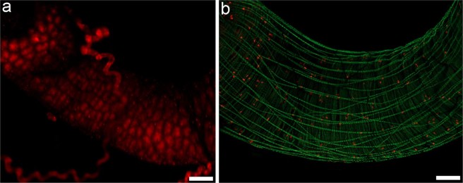
Figure 4.
C. cumulans midgut as an oxidative and mitotically active environment. (a) Midgut labelled with DHE. Analysis under a confocal microscope employing the “z-stack” function and overlap of 350 µm. (b) Midgut mitotic cells revealed by anti-pHH3 antibody (red) and Alexa Fluor 488 phalloidin (green) highlighting muscle fibers. Analysis under a fluorescence stereomicroscope employing the “z-stack” function and overlap of 350 µm. Bars = 100 μm.

