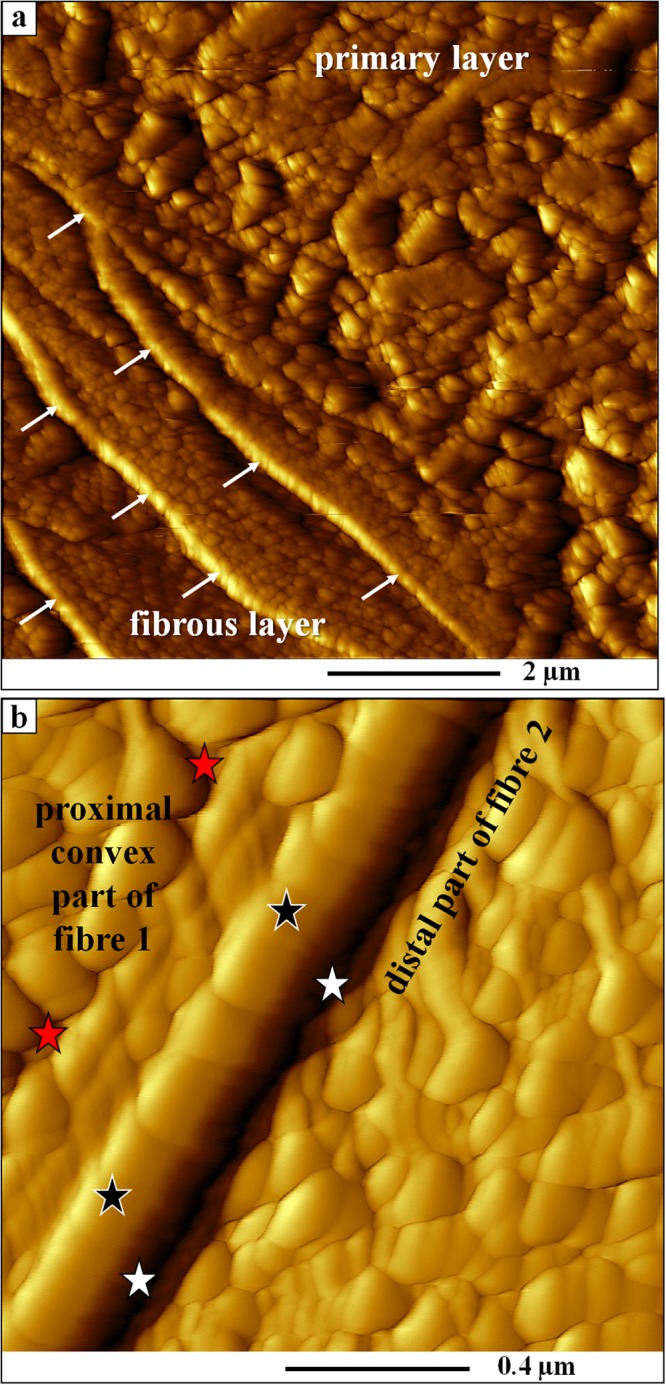
Figure 3.

AFM vertical deflection images depicting the internal structure of primary and fibrous shell portions of Magellania venosa. Corresponding lateral deflection images are shown in Fig. A2. (a) Close-up of the primary layer and the first three rows of adjacent fibres visualizing the gradual changeover from primary to fibrous calcite shell layers. (b) Biopolymer membrane tightly attached to the calcite of a fibre along its proximal, convex surface. The organic membrane (black stars) is between two adjacent fibres (red and white stars) and in each case the biopolymer lines the basal (proximal), convex portion of the fibre.
