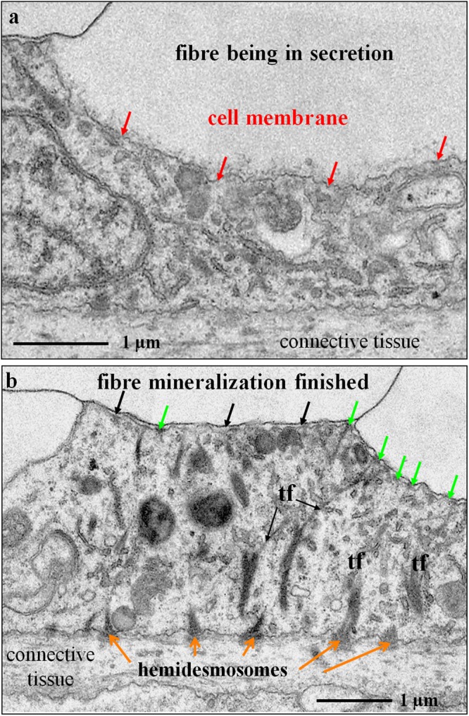
Figure 6.

TEM micrographs of chemically fixed and decalcified contact between epithelium and fibre calcite in modern Magellania venosa. Samples were taken from the central region of the shell. (a) With ongoing mineralization, the membrane lining the proximal, convex part of the fibre is not yet developed (red arrows). (b) Apical cell membrane attached to organic membranes of the fibres by apical hemidesmosomes (green arrows), the latter being connected to basal hemidesmosomes (orange arrows) via tonofilaments (tf). Cells below fibres in the process of active mineral secretion do not contain any tonofilaments.
