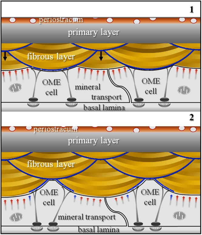
Figure 8.

Schematic model illustrating calcite fibre shape formation for terebratulide and possibly rhynchonellide brachiopods. We see a stack of transversely cut fibres. Prior to fibre secretion OME cell membranes are in close contact with the extracellular organic membrane lining present along the proximal surface of fibres. Detachment of epithelial cells from this membrane lining induces mineral secretion and starts fibre growth (black arrows in schematic 1). When fibres have reached their full width, OME cells start to secrete the organic membrane lining (blue arrows in schematic 2), and when finished, will completely line the basal convex part of the fibre (blue stars in schematic 2).
