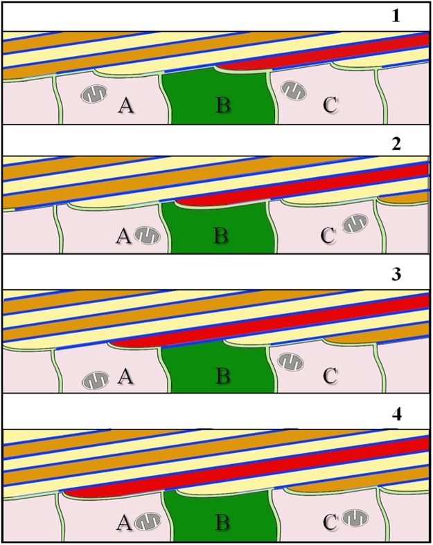
Figure 9.
Schematic model illustrating calcite fibre elongation for terebratulide and rhynchonellide brachiopods. A stack of longitudinally cut fibres is shown. Fibre growth occurs through the coordinated interaction of neighbouring cells (A to C). These are stationary and secrete both, the organic basal lining as well as the calcite of the fibre, and this in the required proportion necessary for the developing fibre (stages 1 to 4). Elongation of fibres takes place by repeated continuous changes in the position of growing fibres relative to cells: (i) attached either to the organic membrane lining the convex surface of the fibre or (ii) to the calcite of the fibre. The organic membrane lining the fibre is indicated with blue lines. Due to the absence of a one-by-one relationship between epithelial cells and calcite fibres, as the shell grows in thickness, each epithelial cell contributes to the formation of more than one fibre and secretes both calcium carbonate and organic material at different portions.

