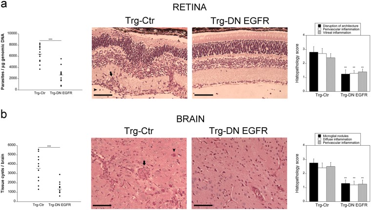
Figure 1.
Expression of DN EGFR diminishes T. gondii load in the eye and brain and enhances resistance to ocular and cerebral toxoplasmosis. Trg-Ctr and Trg-DN EGFR mice were infected with tissue cysts of the ME49 strain of T. gondii and euthanized at 14 days. (a) Retinal levels of T. gondii B1 gene were examined using qPCR. 12 mice per group pooled from 3 independent experiments. Eyes from infected Trg-Ctr showed disruption of retinal architecture, perivascular (arrow) and vitreal inflammation (arrowhead). H&E; X200. Bar, 100 μm. Histopathologic changes in the retina and brain were scored using previously described criteria (refs 33,34). Bars are mean ± SEM of 12 mice per group from 3 pooled experiments. (b) T. gondii tissue cysts per brain. 12 mice per group pooled from 3 independent experiments. Brains from Trg-Ctr mice showed more prominent microglial nodules (arrow) and frequent tissue cysts (arrowhead). PASH original magnification x200. Bar, 100 μm. **p < 0.01, ***p < 0.001 (Student’s t test; Mann-Whitney U test).

