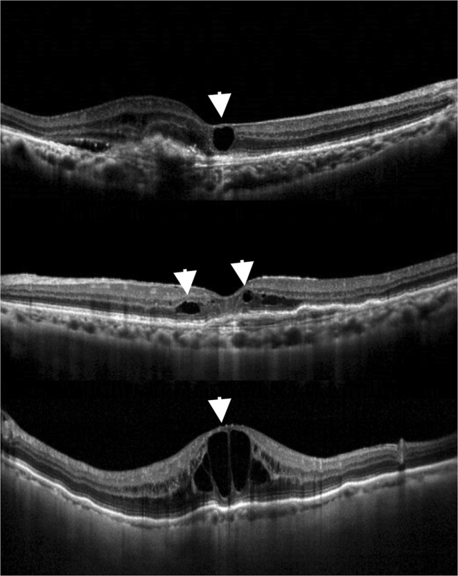
Figure 5.

Optical coherence tomography (OCT) images of different types of morphology of intraretinal cysts (IRCs) in age-related macular degeneration. (Top) OCT image showing degenerative morphology of an IRC (arrowhead) with square-shaped contour and alteration of underlying retinal pigment epithelium. (Middle) Another example of degenerative morphology of IRCs (arrowheads) on OCT, demonstrating a small cyst without obvious expansion of the adjacent layered retinal structure (arrowhead on the right) and an IRC with square-shaped contour (arrowhead on the left). (Bottom) Example of exudative IRCs on OCT, showing large cysts along with stretched adjacent retinal tissue and normal retinal pigment epithelium.
