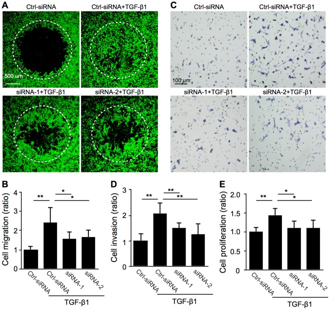
Figure 4.
SNAIL is required for TGF-β1-induced cell motility in Müller glial cells. (A–E) Control siRNA-treated (Ctrl-siRNA) and SNAI1-knockdown (siRNA-1 and -2) Müller glial cells were stimulated with or without TGF-β1 at 10 ng/ml. (A,B) Cell migration assay. Representative images of cells stained with calcein AM. (A) The signal intensity of stained cells having migrated within the detection zones (white dotted circles) after 48 hours was measured (B). n = 7–10 per group. Scale bar = 500 μm. (C,D) Cell invasion assay. Representative images of cells stained with Toluidine Blue O (C). The number of stained cells having invaded on the bottom side of Matrigel-coated filter after 24 hours was counted (D). n = 3 per group. Scale bar = 100 μm. (E) Cell proliferation assay. The optical density for BrdU incorporation after 24 hours was measured. n = 8 per group, *p < 0.05, **p < 0.01.

