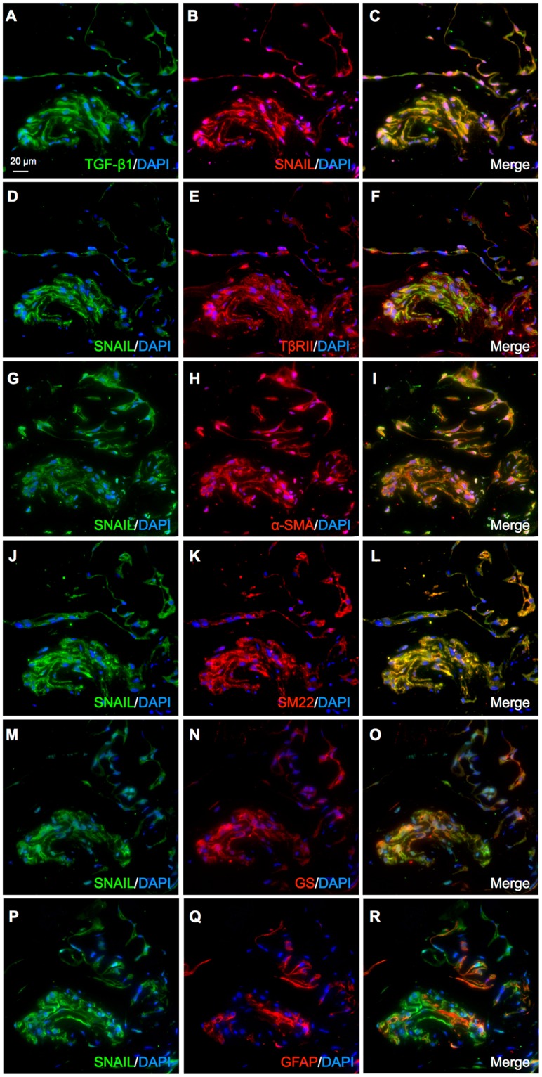
Figure 6.

SNAIL co-localizes with TGF-β ligand-receptor system in cells positive for myofibroblastic and Müller glial markers in iERM patient specimens. (A–O) Double labeling of TGF-β1 (green) and SNAIL (red) (A–C), SNAIL (green) and TβRII (red) (D–F), SNAIL (green) and α-SMA (red) (G–I), SNAIL (green) and SM22 (red) (J–L), SNAIL (green) and GS (red) (M–O), and SNAIL (green) and GFAP (red) (P–R) in the iERM tissue specimens with DAPI (blue) counterstain to nuclei. Scale bar = 20 μm.
