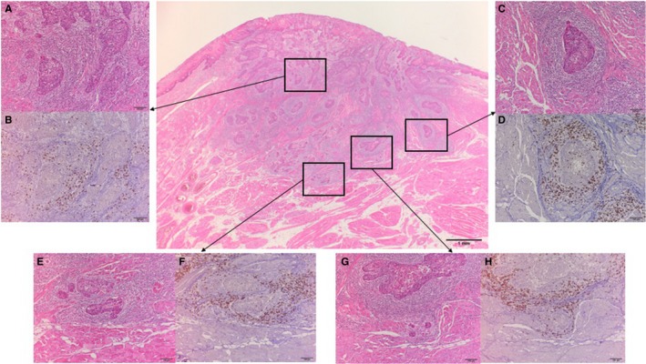
Figure 1.

Representative hematoxylin and eosin (H&E) and CD8 immunohistochemical (IHC) staining of oral squamous cell carcinoma sections for assessment of CD8+ T‐cell density at five different anatomic locations; the parenchyma and stroma in the center of the tumor (A, H & E; B, IHC), the parenchyma and stroma in the invading tumor edge (C, H & E; D, IHC), and the periphery of the tumor (E, H & E; F, IHC; G, H & E; H, IHC). The invading edge is a belt zone including a tumor nest layer inside the tumor border. The periphery of the tumor is outside of the tumor border. Evaluation of peripheral CD8+ T‐cell density included the area of most scattered cancer cells or small islands (G and H rather than E and F). The regions in the rectangle (A, C, E, and G) are shown at ×100 magnification of the arrowed panel
