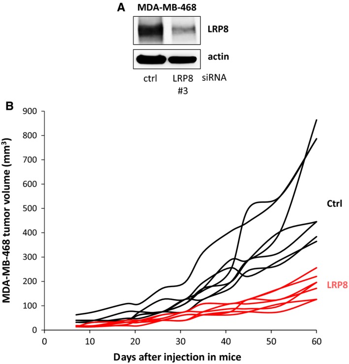
Figure 6.

LRP8 depletion slows tumor growth. MDA‐MB‐468 cells were transfected with control (ctrl, black) or LRP8 (#3, red) siRNAs. A, The level of LRP8 protein was evaluated by Western blotting 24 h after transfection. Actin was used as a loading control. B, Twenty‐four hours after transfection, 4 × 10⁶ MDA‐MB‐468 cells were injected subcutaneously into Swiss nude mice (7 animals/group). Tumor growth was evaluated twice weekly for 2 mo. The data shown are the tumor volume measured for each animal. The differences in tumor volume between the control siRNA and LRP8 siRNA groups were evaluated at each time point, in Wilcoxon tests. We used Benjamini‐Hochberg correction to adjust for multiple testing. Differences were considered significant if the adjusted P value was below 0.05. This was the case for all time points from day 11 (adjusted P = 0.003)
