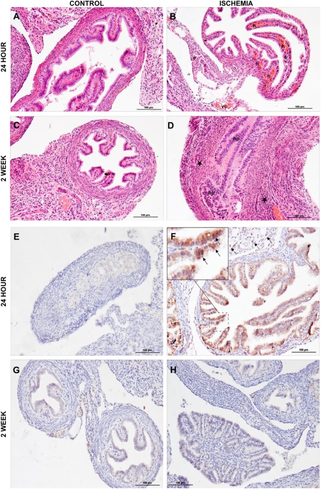
Figure 3.
Histological and immunohistochemical analysis. Histological studies showed that (A) no pathologic changes were detected in control animals at 24 hours postischemia. B, Vascular congestion, edema, and severe intraparenchymal hemorrhage were observed in ischemia and reperfusion (IR) group at 24 hours postischemia. C, Normal histology was observed in the control group at 2 weeks postischemia. D, Increased inflammatory cell infiltration (the majority are leucocytes and macrophages) and epithelial cell proliferation determined at 2 weeks postischemia. Vascular congestion (vc), hemorrhage (h), edema (e), hyperplasia (hp), and inflammatory cells infiltration (*). Immunohistochemical studies showed (E) caspase 3 expression in the control group at 24 hours after ischemia. F, Increased caspase-3 expression was observed in epithelial (dashed arrow in magnified view) and stromal (arrow) cells of IR group at 24 hours after ischemia. G, Caspase-3 expression in the control group 2 weeks after ischemia. H, Caspase-3 expression gradually decreased and comparable levels with the control group were seen at 2 weeks after ischemia.

