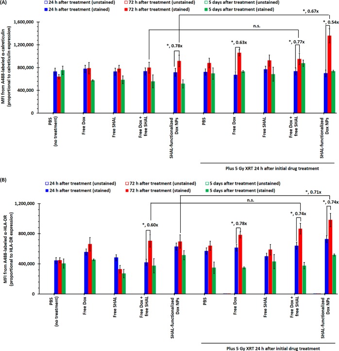
Figure 5.
Calreticulin and HLA-DR expressions of Raji cells after treatment with a subtherapeutic dose (i.e., IC25) of free/encapsulated Dox with/without a therapeutic dose of X-ray irradiation. (A) The plot of mean fluorescence intensities (MFI) of unstained and α-calreticulin-stained Raji cells after treatment with IC25 of either free or encapsulated Dox, with or without 5 Gy X-ray irradiation (24 h after the initial drug treatment). (B) The plot of mean MFI of unstained and α-HLA-DR-stained propidium iodide-negative (PI–) variable Raji cells after treatment with IC25 of either free or encapsulated Dox, with or without 5 Gy X-ray irradiation (24 h after the initial drug treatment). Both antibodies were A488 stained (N.B., n = 3; * denotes p < 0.05, and hence statistically significant).

