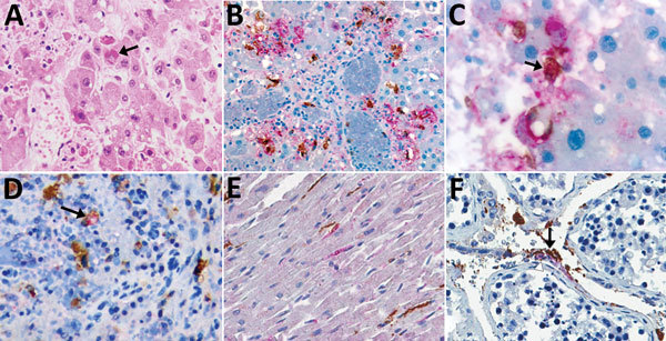
Figure 2.

Immunohistochemical stains of tissue from patients with fatal cases of Ebola virus (EBOV) disease showing EBOV (red) and CD163 (brown) antigens. A) Hematoxylin and eosin stain of liver showing hepatocellular necrosis with intracytoplasmic eosinophilic inclusions (arrow). B) EBOV antigens in hepatocytes and CD163 antigens in macrophages. C) High magnification image of double immunohistochemical staining of liver tissue showing colocalization of EBOV and CD163 antigens in macrophage (arrow). D) Colocalization of EBOV and CD163 antigen in macrophage of spleen (arrow). E) Staining of EBOV and interstitial macrophages (CD163) in heart. EBOV found in some cardiomyocytes. F) EBOV and CD163 antigen in endothelial cells (arrowhead) and macrophages of testis (arrow). Original magnification ×20 (A, B, D, E, F); ×63 (C).
