Figure.

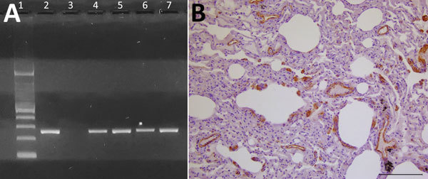
Evidence of dolphin morbillivirus infection in Eurasian otters (Lutra lutra), southwestern Italy. A) Comparison of nucleoprotein gene amplification products from infected otters, obtained by reverse transcription PCR. A specific band at the expected molecular weight of 287 bp is shown. Lane 1, molecular weight marker (Tracklt 100bp DNA Ladder; Invitrogen, http://www.thermofisher.com); lane 2, positive control (lung tissue from an infected striped dolphin, Stenella coeruleoalba); lane 3, negative control (distilled water); lanes 4–7, samples from morbillivirus-positive Eurasian otters: LL-290, lung (lane 4); LL-291, kidney (lane 5); LL-3380, lung (lane 6); LL-7318, lung (lane 7). B) Mayer’s hematoxylin counterstain of lung tissue shows marked and widespread immunohistochemical labeling for morbillivirus antigen (dark areas), particularly evident at the level of vascular walls and endothelial cells and, to a lesser extent, of alveolar epithelial cells (morphologically consistent, or not, with hyperplastic type II pneumocytes) as well as of thickened alveolar septa. Scale bar indicates 100 μm.
