Fig. 2.
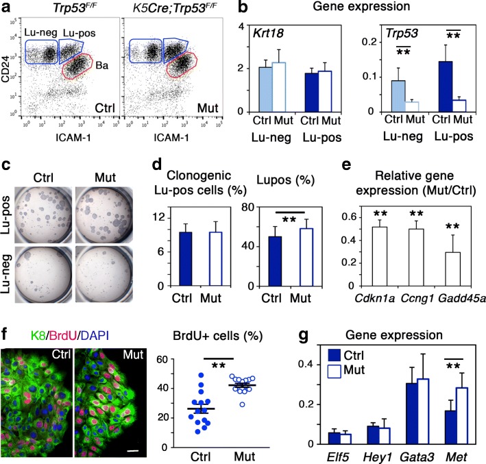
Loss of p53 induces expansion of luminal progenitors overexpressing Met. a Flow cytometry analysis of CD24 and ICAM-1 expression in mammary cells isolated from Trp53F/F (Ctrl, control) and K5Cre;Trp53F/F (Mut, mutant) adult virgin mice. b qPCR analysis of Krt18 and Trp53 expression in Lu-neg and Lu-pos cells isolated from control and mutant adult virgin mice. Data are the mean ± SEM of 4 separate cell preparations. **p ≤ 0.01. c Microphotographs of the colonies formed by 500 control and mutant Lu-neg and Lu-pos cells. d Left: Percentages of clonogenic cells in control and mutant Lu-pos cell populations. Data are the mean ± SEM of 3 distinct assays. Right: Percentages of Lu-pos cells in control and mutant luminal cell populations calculated from flow cytometry data. Values shown represent the mean ± SEM of 7 separate analyses of distinct cell preparations. e Expression of Cdkn1a, Ccng1, and Gadd45a (encoding p21, cyclin G1, and Gadd45a, respectively) in Lu-pos cells isolated from control and mutant adult virgin mice, evaluated by qPCR. Data are shown as mean ratios (± SEM) between gene expression levels in mutant and control Lu-pos cells from 4 separate preparations. **p ≤ 0.01. f Left: K8 and BrdU immunodetection in colonies derived from control and mutant Lu-pos cells. DAPI-stained nuclei appear in blue. Bar, 20 μm. Right: Percentages of BrdU+ cells. Each point represents counting of one microscope field comprising 100–300 DAPI-stained nuclei. Data are shown as mean ± SEM of countings from 2 distinct experiments. **p ≤ 0.01. g Expression levels of the luminal-specific regulators, Elf5, Hey1, Gata3, and Met in control and mutant Lu-pos cells, evaluated by qPCR. Data are the mean ± SEM of 3–4 separate preparations. **p ≤ 0.01

