Figure 1.

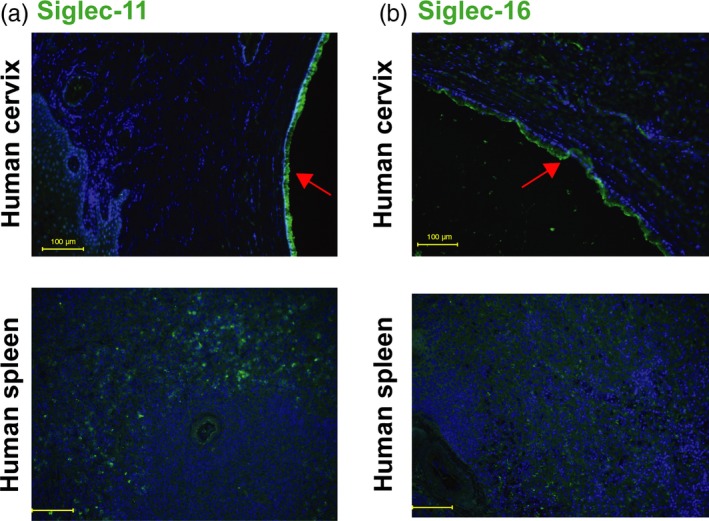
Siglec‐11 and Siglec‐16 are expressed on human cervical epithelium. Immunohistochemical analysis of paraffin sections of human cervix samples, with spleen as positive control using mouse monoclonal. (a) anti‐Siglec‐11 and (b) anti‐Siglec‐16 antibodies (green). Nuclei are blue (Hoechst), red arrows indicate columnar cervical epithelium, and yellow bar indicates 100 µm. Images are representative of n = 3. The samples of human Cervix and spleen shown were heterozygous for SIGLEC16
