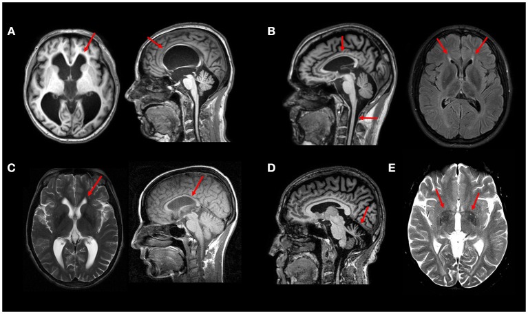
Figure 2.
Typical MRI findings in distinct HSP subtypes—(A) Axial and sagittal T1W images showing hydrocephalus due to aqueductal stenosis in a patient with SPG4. This finding is rather unusual in SPG4, but relatively frequent in SPG1 patients. (B) Sagittal T1W image showing thin corpus callosum and spinal cord atrophy in a patient with SPG11; Axial FLAIR image showing the “ear-of-the-lynx” sign in the same patient. (C) Thin corpus callosum (T1W image) and subtle “ear-of-the-lynx” sign (T2W image) in a patient with SPG15. (D) Sagittal T1W image revealing marked cerebellar atrophy in a patient with SPG7. (E) White matter hyperintensities and T2 hypointense signal at the globi pallidi in a 21-years old patient with SPG35 (T2W image). All images were obtained during routine clinical care of these patients at UNICAMP and UNIFESP hospitals.

