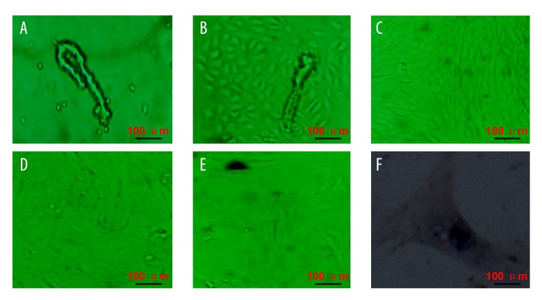
Figure 1.
Isolation and identification of rat brain microvessel endothelial cells. (A) Isolated microvessel segments and/or single cells. (B) Short spindle and/or polygonal cells were observed around microvessel fragments. (C) Paving stone-like cells. (D) Monolayer growth endothelial cells. (E) Passaged rat brain microvessel endothelial cells. (F) Immunocytochemistry identified brain microvessel endothelial cells. Scale bars are illustrated in figures.

