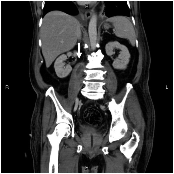
Figure 2.

A computed tomography image of the abdomen (coronal view) during the 2nd week of hospitalization shows a cystic lesion approximately 3.3 cm in size over the right psoas muscle with wall enhancement (white arrowhead), which could not be drained via the percutaneous approach
