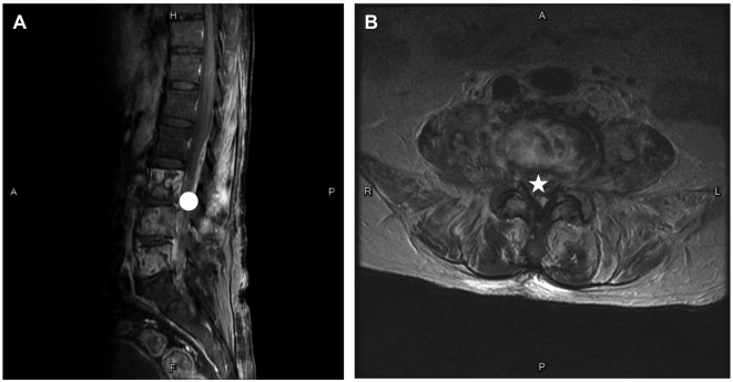
Figure 3.
Images taken on the patient’s 21st day of hospitalization. (A) Magnetic resonance image of the lumbar spine (sagittal view) shows fluid collection in the L3/4 and L4/5 intervertebral disc spaces together with narrow enhancement of the L3–L5 vertebral bodies (circle). (B) Magnetic resonance image of the lumbar spine (axial view) shows abnormally enhanced lesions involving the bilateral psoas muscles, prevertebral space and intraspinal canal (star)

