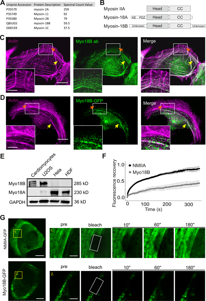
Figure 1. Myosin-18B is a stable component of contractile stress fibers.

(A) Top myosin family protein hits from the BioID screen for identification of tropomyosin 1.6 (Tpm1.6) -associated proteins in U2OS cells. (B) Domain structures of myosin IIA, myosin-18A and myosin-18B. All three proteins contain a motor-like head domain followed by the coiled-coil (CC) region. Myosin-18A additionally contains a lysine/glutamate (KE) -rich region and the PDZ domain in its N-terminal extension, whereas myosin-18B harbors large N- and C-terminal extensions with unknown domain structures. (C) Immunofluorescence microscopy analysis demonstrating that in U2OS cells endogenous myosin-18B localizes to F-actin -rich (visualized by fluorescent phalloidin) ventral stress fibers (indicated by yellow arrows), but not to non-contractile dorsal stress fibers (indicated by orange arrows). (D) Also myosin- 18B-GFP fusion protein co-localizes with myosin IIB to ventral stress fibers. Bars, 10 µm and 5 µm in the panels C, D and magnified insets, respectively. (E) Western blot analysis of endogenous myosin- 18B and myosin-18A in U2OS, HeLa, and HDF cells. GAPDH was probed for equal sample loading. Please note that the doublet band in the cardiomyocyte extract at ~280 kDa may result from post- translational modification of myosin-18B. (F) Normalized average Fluorescence-recovery-after- photobleaching (FRAP) recovery curves of NMIIA (black line) and myosin-18B (gray line) demonstrating their dynamics on stress fibers. 23 myosin-18B-GFP transfected cells and 22 NMIIA- GFP transfected cells were used for quantification. The data are presented as mean ± s.e.m. (G) FRAP analysis of the dynamics of NMIIA-GFP and myosin-18B-GFP in contractile stress fibers of U2OS cells. Examples of cells expressing NMIIA-GFP and myosin-18B-GFP are shown on the top, and magnified regions on the bottom represent time-lapse images of the bleached regions. Bars, 10 µm (in cell images) and 2 µm (in the magnified time-lapse images). See also Figure S1 and Table S1.
