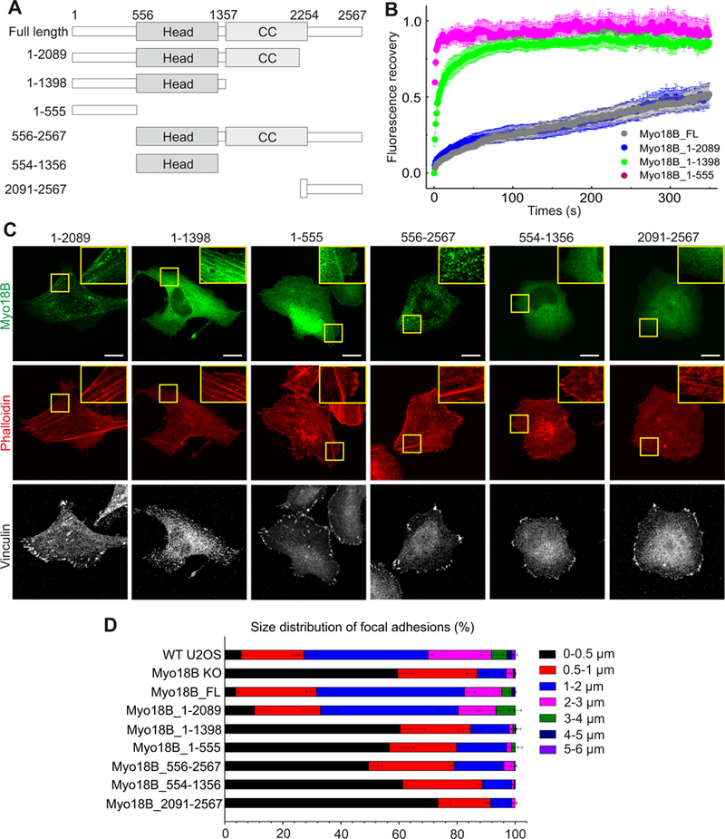
Figure 7. Localization and dynamics of myosin-18B deletion constructs, and their effects on stress fiber and focal adhesion maturation.

(A) Domain structures of GFP-tagged myosin-18B constructs used in the FRAP and rescue experiments. (B) Normalized average FRAP recovery curves of different myosin-18B-GFP constructs expressed in myosin-18B knockout U2OS cells. 23 myosin-18B-FL-GFP, 18 myosin-18B-1-2089-GFP, 12 myosin-18B-1-1398-GFP, and 13 myosin-18B-1-555-GFP transfected cells were used for quantification. The data are presented as mean ± s.e.m. (C) Representative images of myosin-18B knockout cells transfected with different myosin-18B-GFP constructs shown in panel A. F-actin and focal adhesions were visualized by phalloidin and an antibody against vinculin, respectively. Magnified regions (corresponding to the yellow boxes) highlight localization of the proteins to stress fibers. Bars, 10 µm. (D) The length distributions of vinculin-positive focal adhesions. The number of focal adhesions in each size group was divided with the total adhesion number of the same cell. n = 988 adhesions from 14 cells (wild-type); 2520 adhesions from 16 cells ( myosin-18B knockout,); 979 adhesions from 15 cells (myosin-18B-FL); 1236 adhesions from 12 cells (myosin-18B- 1–2089); 1738 adhesions from 13 cells (myosin-18B-1–1398); 1404 adhesions from 13 cells (myosin18B-1–555 rescue); 2470 adhesions from 13 cells (myosin-18B-556–2567); 1495 adhesions from 13 cells (myosin-18B-554–1356); 1671 adhesions from 13 cells (myosin-18B-2091–2567). Data are represented as mean ± s.e.m. See also Figures S6 and S7.
