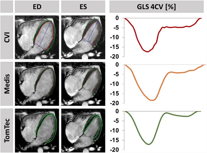
Fig 1. Feature-tracking using different software solutions.
On the left, endo- and epicardially tracked borders of the left ventricle in a 4 chamber view (CV) at the end-diastole (ED) and end-systole (ES) are shown in a healthy volunteer using the different commercially available software solutions (upper row: CVI, middle row: Medis, bottom row: TomTec). On the right, the corresponding global longitudinal strain (GLS) curves are displayed.

