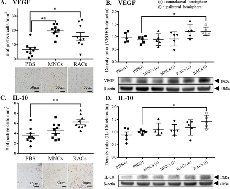
Fig 5. Immunohistochemistry and western blotting of ipsilateral hemisphere tissue at day 7.
(A) Injection of either MNCs or RACs significantly increased VEGF-positive cells in the peri-infarct area at day 7 (each group: n = 9) (**P<0.01 and *P<0.05, respectively, Kruskal-Wallis test). Scale bar: 50 μm. (B) Injection of RACs (1 x 10⁴/50 μL) significantly increased VEGF in the peri-infarct area, as determined by western blotting, at day 7 (*P<0.05, Kruskal-Wallis test). (PBS: n = 6, MNCs: n = 6, RACs: n = 7) (C) Injection of RACs (1 x 10⁴ /50 μL) significantly increased IL-10-positive cells in the peri-infarct area at day 7 (*P<0.05, Kruskal-Wallis test) (each group: n = 9). Scale bar: 50 μm. (D) Injection of RACs (1 x 10⁴ /50 μL) significantly increased IL-10 in the peri-infarct area, as determined by western blotting, at day 7 (*P<0.05, Kruskal-Wallis test) (PBS: n = 6, MNCs: n = 6, RACs: n = 7).

