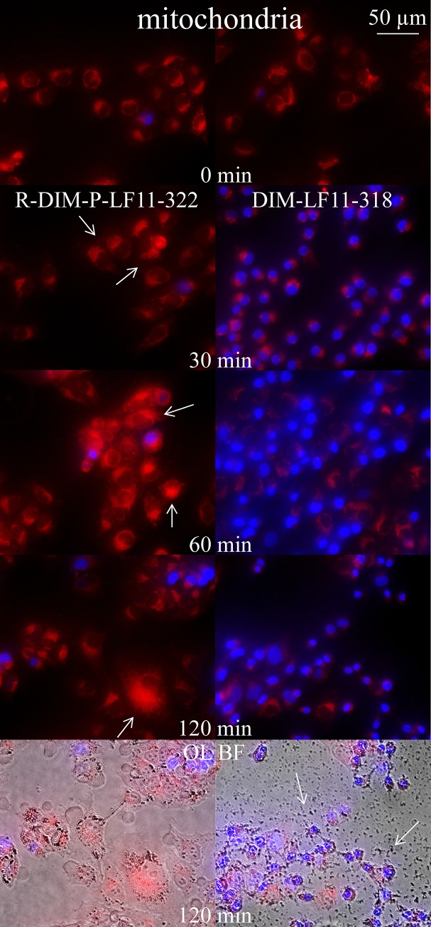
Fig 11. Changes in morphology of mitochondria of A375 upon incubation with R-DIM-P-LF11-322 and DIM-LF11-318.
Overlay images of melanoma cells (A375) with stained mitochondria (red, MitoTracker Deep Red) and nucleus (blue, NucBlue) in presence of 10 μM peptide R-DIM-P-LF11-322 (left) or DIM-LF11-318 (right) at different incubation periods of 0, 30, 60 and 120 min. R-DIM-P-LF11-322 causes significant mitochondrial swelling (arrows), whereas DIM-LF11-318 mainly induces cell death indicated by increased uptake of nuclear staining with no significant swelling of mitochondria. Bottom OL BF: Overlay of bright field (OL BF) and respective fluorescence. Arrows indicate membrane debris upon incubation with DIM-LF11-318. Pictures are representative for a series of three experiments.

