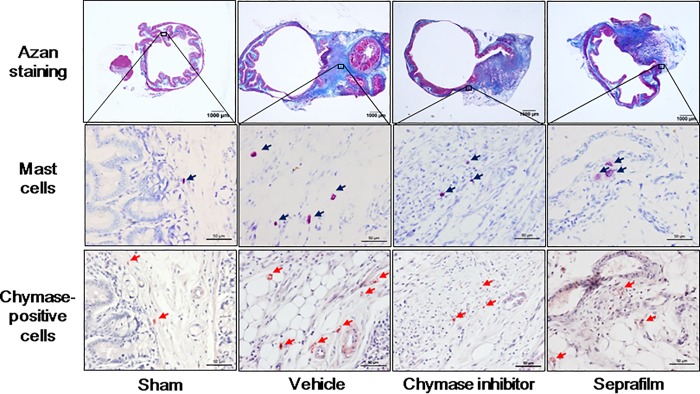
Fig 7. Mast cells and chymase-positive positive cells in cecum.
Representative images of cecal sections stained with Azan-stain and toluidine blue (mast cells), and immunostained with anti-chymase (chymase-positive cells) in the sham-, vehicle-, chymase inhibitor-, and Seprafilm-treated rats 2 weeks after alkali-injury. The original magnification is 20X (upper images); scale bars are 1 mm. The original magnification is 200X (middle and lower images); scale bars are 0.05 mm.

