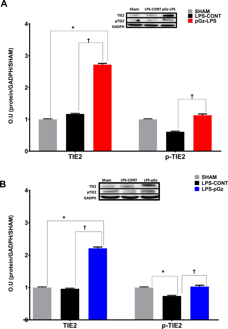
Fig 3. Expression and phosphorylation of TIE2 in mice after LPS.
Expression of the tyrosine kinase receptor tunica interna endothelial cell kinase 2 (TIE2) and its phosphorylation in mice, 6 hrs. after LPS. LPS did not significantly change expression of TIE2, but significantly decreased TIE2 phosphorylation. (A) pGz pre-treatment (pGz-LPS) and (B) pGz post-treatment (LPS-pGz) significantly increased both TIE2 expression and phosphorylation. Inserts are representative western blots of protein expression of TIE2, p-TIE and GADPH in Sham, LPS-Control, and pGz-LPS and LPS-pGz. LPS-CONT (n = 8) pGz-LPS (n = 8), LPS-pGz (n = 8), or Sham (received equal volume of phosphate buffer but did not received LPS or pGz, n = 4). Data are mean ± SEM (* p<0.01 vs Sham, Ɨ p<0.01 vs. LPS-CONT).

