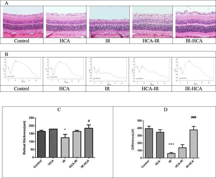
Fig 2.
(A) Hematoxylin and eosin staining of the retinas. Representative photomicrographs showing the histologic appearance of retinas in the five groups. Significant decrease in the mean retinal thickness is visible in the IR group. (B) Representative ERG waveforms from the five groups. I/R injury significantly reduced the the α and β wave amplitudes. (C)Whole retinas were significantly thicker in the IR-HCA group than in the IR group. (D) ERG revealed significant recovery in the difference of cursor of α and β waves in the IR-HCA group. The results are mean ± SEM of three independent experiments. ***P < 0.001, *P < 0.05 versus control group; ###P < 0.001, #P < 0.05 versus IR group.

