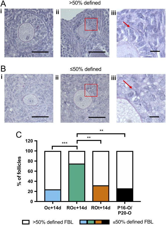
Figure 5.

Analysis of basal lamina in ovaries and reaggregated ovaries. (Ai and ii) Representative images of follicles stained with Periodic acid-Schiff and haematoxylin defining how follicle basal lamina (FBL) was classified as >50% defined. (Aiii) Higher magnification of the red box in Aii, with the red arrow pointing at the stained FBL. (Bi, ii) Representative images of follicles classified as ≤50% defined FBL. (Biii) Higher magnification of the red box in Bii, with the red arrow pointing at the lack of FBL definition. (C) Analysis of FBL definition in primary, secondary and preantral follicles in cultured ovaries (Oc + 14d), cultured reaggregated ovaries (ROc + 14d) and transplanted reaggregated ovaries (ROt + 14d) and in vivo Control ovaries (P16-O/P20-O). Scale bar (Ai, Aii, Bi and Bii): 50 µm; high magnification of FBL scale bar (Aiii and Biii): 10 µm. P16-O/P20-O n = 23 follicles, n = 4 ovaries; Oc + 14d n = 25 follicles, n = 4 ovaries; ROc + 14d n = 24 follicles, n = 3 ovaries; ROt + 14d n = 19 follicles, n = 4 ovaries. **P ≤ 0.01; ***P ≤ 0.001.

 This work is licensed under a