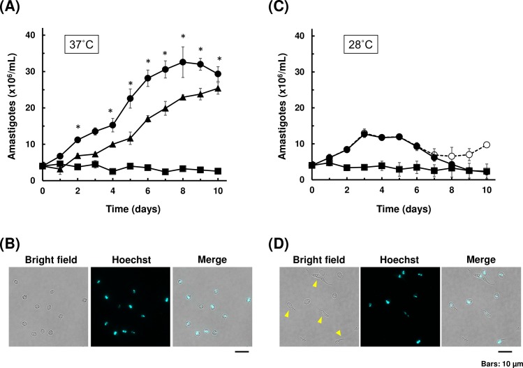
Fig 1. Growth and morphology of axenic amastigote culture.
(A) Growth curves of axenic amastigotes at 37°C. Amastigotes derived from in vitro amastigogenesis were cultured in either LIT medium (closed circle) or DMEM (closed square) at 37°C. Intracellular amastigotes isolated from host 3T3 cells were also cultured in LIT medium (closed triangle). The number of amastigotes were counted under microscopy. At least two independent experiments were performed in triplicates for each group, and mean values (±SD) of one representative experiment were plotted. Significant difference compared to DMEM was indicated (*p<0.05, Kruskal-Wallis test). (B) Morphology of amastigote after 10 days of axenic cultivation in LIT medium at 37°C. The parasites were fixed and stained using Hoechst 33342 to detect nucleus and kinetoplast. Scale bar: 10 μm. (C) Growth curves of axenic amastigotes at 28°C. Amastigotes derived from in vitro amastigogenesis were cultured in either LIT medium (closed and open circles) or DMEM (closed square) at 28°C. For LIT 28°C, closed circle represents the number of round form amastigote only, and open circle represents the total number of parasites including intermediate morphologies. At least two independent experiments were performed in triplicates for each group, and mean values (±SD) of one representative experiment were plotted. No significant difference was observed among groups (Kruskal-Wallis test). (D) Representative image of parasites displaying intermediate morphology after 10 days of axenic cultivation at 28°C (open circle in (C)). Parasites with intermediate forms are indicated by arrow heads.

