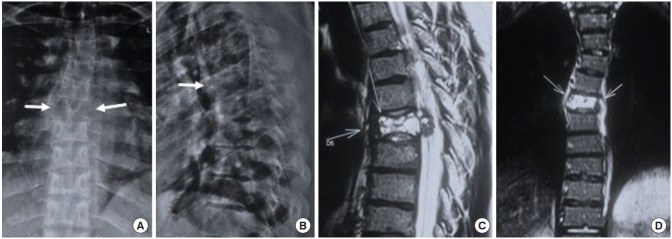
Fig. 1.
(A) Anteroposterior radiograph: arrows point toward loss of pedicle margins on both sides and osteolytic lesion involving the entire D6 vertebra. (B) Lateral radiograph shows mild collapse of the vertebra. (C, D) Sagittal and coronal images of T2-weighted magnetic resonance imaging show an expansile lytic lesion with thinned out sclerotic margin and multiple fluid levels replacing the D6 vertebral body, along with retropulsion and soft tissue expansion of lesion posteriorly into the spinal canal resulting in secondary spinal canal stenosis and cord edema.

