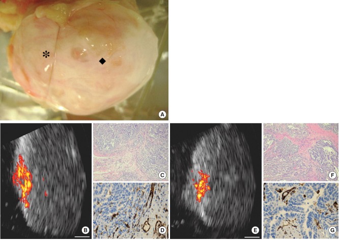
Fig. 10.
Photoacoustic imaging of surgical ovarian specimen. Photoacoustic imaging of surgical ovarian specimen from a 58-year-old postmenopausal patient with bilateral ovarian cancers at stage IIIC. (A) Malignant ovary imaged at 2 locations (* and ◆). (B) Coregistered US and photoacoustic image of location *. (E) Coregistered US and photoacoustic image of location ◆. Highly vascularized intraepithelial areas compared with the surrounding tissue are observed on photoacoustic images of both locations. (C, F) Hematoxylin-eosin stained images (× 40) of the corresponding areas show extensive highgrade tumors. (D, G) CD31-stained images (×100) of the corresponding areas show extensive thin-walled micro vessels. White bar= 5 mm. Reprinted from Aguirre et al[23] Transl Oncol 2011;4:29-37, with figure citation to the Neoplasia Press, Inc.

