
Figure 7. Co-polymerization of GFP:FUS, GFP:FUS tyrosine-to-serine mutants, and GFP:hnRNPA1 to mCherry:FUS seed fibers.
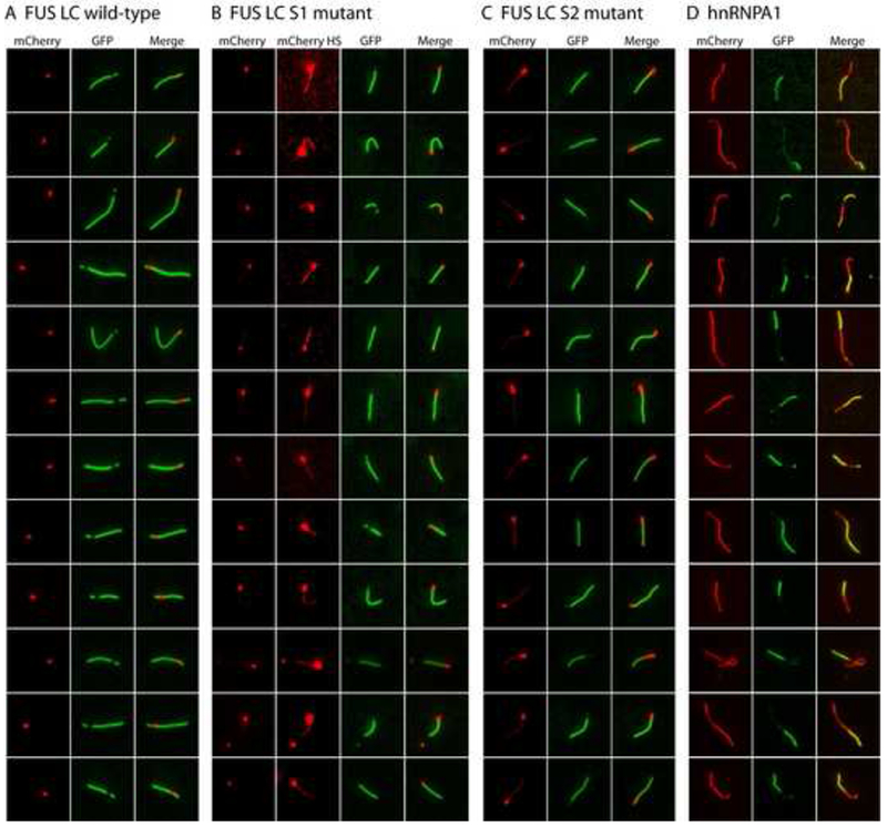
(A) Extensions of pre-formed mCherry:FUS LC fiber seeds by GFP-FUS LC monomers. mCherry:FUS seeds were mixed with monomeric GFP:FUS LC domain (wild-type) at an approximately 5-fold ratio of GFP:FUS to mCherry:FUS seeds. After incubation for 2 hr, fluorescent images for extended fibers were taken in a standard epifluorescent mode on an Olympus TIRF microscope (Experimental Procedures).
(B) Co-extension of mCherry:FUS seeds by monomeric GFP:FUS LC S1 mutant. Higher contrast mCherry images (mCherry HS) revealed that the monomeric S1 mutant (green color) co-polymerized with mCherry:FUS monomer from the seeds.
(C) Co-extension of mCherry:FUS seeds by monomeric GFP-FUS S2 mutant. mCherry signals in the co-polymerization region was stronger than that of (B) and visible in standard contrast images.
(D) Heterotypic co-polymerization of mCherry:FUS and GFP:hnRNPA1. mCherry:FUS seeds were mixed with monomeric GFP:hnRNPA1 LC at equal ratios. Red regions are interpreted to be mCherry:FUS seeds that extended homomeric polymerization during the reaction, green regions are interpreted to reflect heteromeric polymerization of GFP:hnRNPA1 into the mCherry:FUS seeds. Merged images reveal regions of yellow color interpreted to reflect co-polymerization of mCherry:FUS and GFP:hnRNPA1. Additional examples of heterotypic co-polymerization were provided in Figure S5.