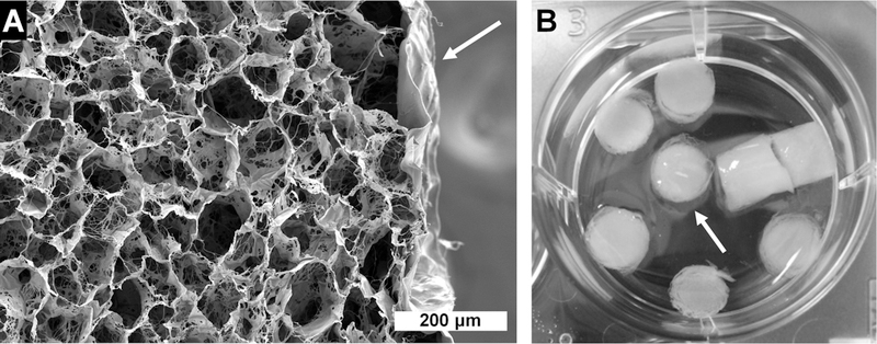
Figure 3.
Images of amniotic membrane-wrapped scaffolds. (A) Scanning electron microscopy of core–shell scaffolds with a collagen–chondroitin sulfate core and amniotic membrane shell (white arrow). The scale bar is 200 μm. (B) Macro image showing hydrated scaffolds in the well of a 6-well plate; the amniotic membrane indicated by a white arrow.

