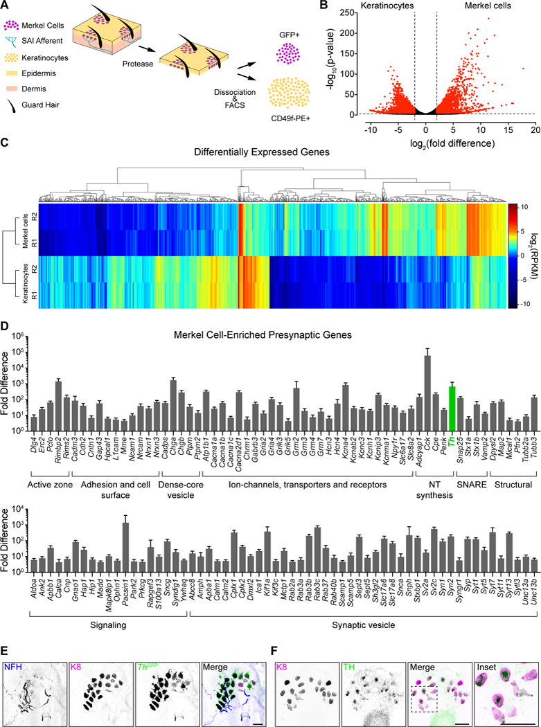
Figure 1. Merkel cells are presynaptic, catecholaminergic cells.

A. Epidermal cell purification strategy [n=2 fluorescence-activated cell sorting (FACS) purifications from n≥2, 7–8-week Atoh1GFP mice each]. B. Volcano plot of genes differentially expressed between Merkel cells and keratinocytes. Dashed lines indicate log2(fold difference)≥2 and Padj<0.01 for differential expression (red, above threshold; black, below threshold). C. Hierarchical clustering of differentially expressed genes. Rows represent RNA-seq replicates (R1/R2). Dendrograms show expression profiles of genes (top) and replicates (left). RPKM, reads per kilobase of exon per million reads mapped. Genes with RPKM<2 across all samples are not displayed. D. Presynaptic genes enriched in Merkel cells are grouped according to functional class. Log2 transformed fold difference is plotted. E. Axial projection of a whole-mount touch dome from an adult ThGFP mouse stained with antibodies against NFH (blue in merge), K8 (magenta) and GFP (green). F. Maximum projection of a touch dome in an epidermal peel stained with antibodies against K8 (magenta) and TH (green). Scale bars, 25 μm. See also Figure S1 and Tables S1 and S2.
