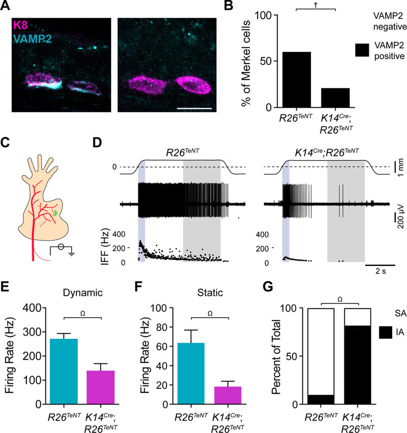
Figure 3. Merkel cells employ SNARE-dependent vesicular release to mediate SAI responses.

A. Images of Merkel cells (magenta) and VAMP2 (cyan) immunoreactivity from littermate-control (R26TeNT) and TeNT-expressing mice (K14Cre;R26TeNT; scale bar, 10 μm). B. Quantification of Merkel cells with detectable VAMP2 immunoreactivity (cyan; two-sided Fisher’s exact test, †P<0.0001; n=149–161 cells from 3 mice per group). C. Schematic of ex vivo skin-nerve recording preparation (saphenous nerve, red; FM1–43-labeled touch dome, green). Receptive fields of touch-dome afferents were identified by FM1–43 fluorescence in epidermal-side up recordings. D. Recordings from touch-dome afferents from littermate-control (left) and K14Cre;R26TeNT (right) mice. Top traces, displacement (dashed lines, point of skin contact). Middle traces, action potential trains. Bottom, instantaneous firing frequency (IFF) plots [blue region, dynamic (ramp) phase; gray region, static (late hold) phase]. (E–G). Maximal touch-evoked responses from littermate-control (cyan) and K14Cre;R26TeNT (magenta). Peak dynamic firing rate (E) and mean static firing rate (F). Mean±SEM; two-tailed Mann-Whitney test, Ω.P<0.005. G. Units were classified as slowly adapting (SA, white) if spikes were observed throughout the 5-s hold phase in ≥75% of stimulus presentations, otherwise they were classified as intermediately adapting (IA, black; two-sided Fisher’s exact test, ΩP<0.005; n=10–11 fibers, 9 mice per group). See also Figures S3 and S4.
