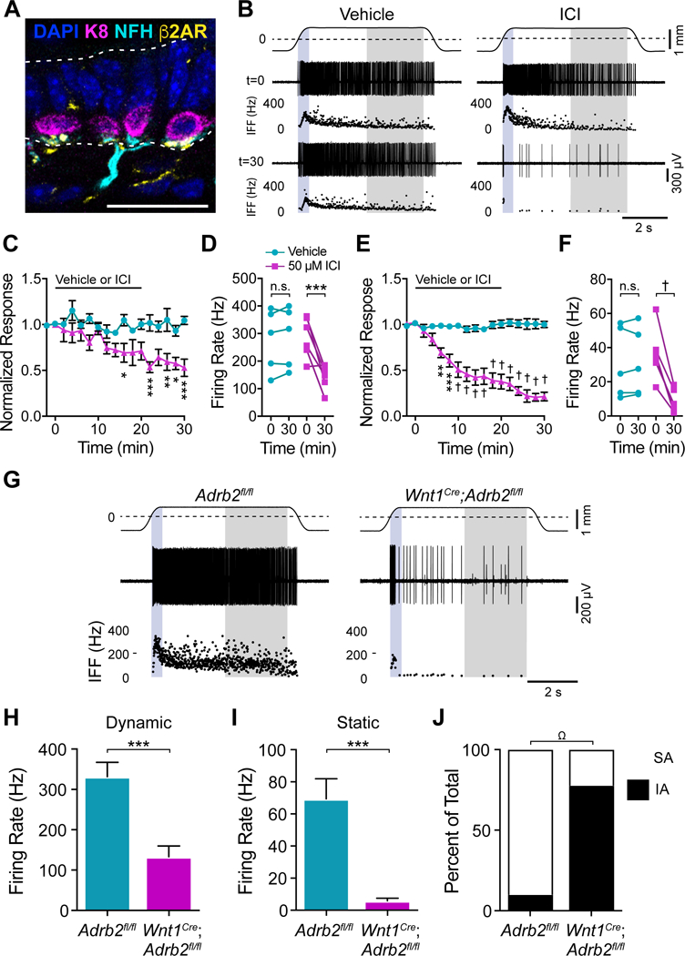
Figure 5. Neuronal β2ARs are required for touch-evoked SAI responses.

A. Merkel cells (K8, magenta), Merkel-cell afferent (NFH, cyan), and β2AR (yellow) immunoreactivity in adult mouse back skin (scale bar, 25 μm). B. Recordings from Merkel-cell afferents and corresponding IFF plots before and 30 min after treatment with vehicle or 50 μM ICI 118,551. Traces and shading as in Fig. 3D. (C–F). Peak dynamic firing rate (C, D) and mean static firing rates (E, F) from afferents treated with vehicle (cyan) or 50 μM ICI 118,551 (magenta) and stimulated at 2-min intervals. C, E. Firing rates at each time point was normalized to t=0 to average responses across units (mean±SEM). Black bar indicates time of vehicle or 50 μM ICI 118,551 application. D, F. Firing rates of all units before (t=0) and after (t=30) treatment with vehicle or 50 μM ICI 118,551. Two-way matched ANOVA with Sidak’s post-hoc; *P<0.05, **P<0.01, ***P<0.001, †P<0.0001; n=5–6 units from 5–6 mice per group. G. Recordings from Merkel-cell afferents from littermate control (left) and Wnt1Cre;Adrb2fl/fl (right) mice. (H–J). Maximal touch-evoked responses from littermate-control (cyan) and Wnt1Cre;Adrb2fl/fl (magenta) units. H. Peak dynamic firing rate. I. Mean static firing rate (two-tailed Student’s t test, ***P<0.001; mean±SEM). J. Proportion of Merkel-cell afferents with SA (white) versus IA firing patterns (black); two-sided Fisher’s exact test, ΩP<0.005; n=9–10 units, 9 mice per group). See also Figure S5.
