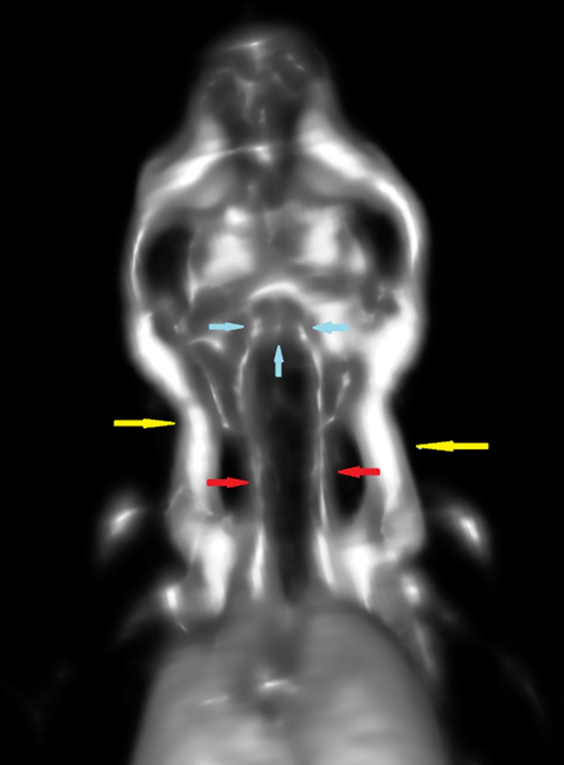
Fig 6. MicroPET images of the mouse head and neck allow visualization of major head and neck vessels.

Major vessels in the neck of the mice injected with FDG-labeled erythrocytes could be visualized, such as the external jugular veins in the lateral neck (yellow arrows), the smaller common carotid arteries in the central neck (red arrows), and the circle of Willis (blue arrows).
