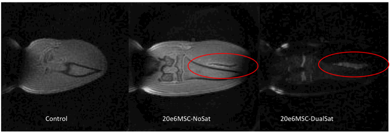
Figure 4.

(A) –SWIFT MR image of a rat knee, (B) - SWIFT MR image of a rat knee after administration of the high concentration of iron labeled MSCs into a muscle tissue. The hypointense signal from grafted cells was detected, (C) – the same image as (B) with tissue saturation pulse. Hyperintense signal from grafted cells was recovered.
