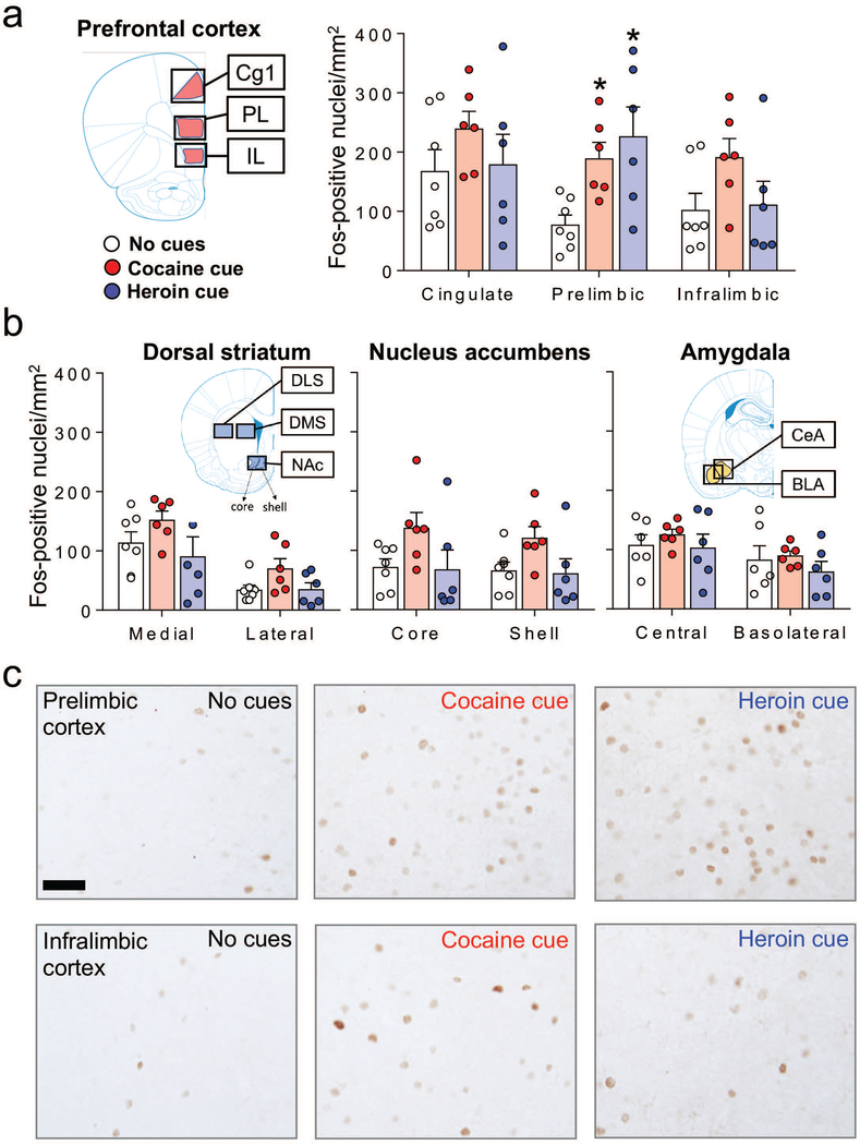
Figure 2.
Cue-induced reinstatement of cocaine or heroin seeking is associated with Fos induction in the PL, but not in the other prefrontal cortical areas (Cg1 and IL), and not in the striatum or amygdala. (a) Number of Fos-positive nuclei/mm2 (mean ± SEM) in mPFC (Cg1, PL and IL subregions), (b) dorsal striatum (medial and lateral subregions), nucleus accumbens (core and shell subregions), and amygdala (CeA and BLA subregions) for the no cues (n = 6–7), cocaine cue (n = 6), and heroin cue (n = 6) groups. *p< 0.05 relative to the no cues group. Images for each brain region were captured from the areas indicated by the outer black boxes on the coronal section schematics. The specific sampling areas used for quantifying Fos-positive nuclei are indicated by the colored overlays. (c) Representative images of Fos-positive nuclei in PL and IL cortex. Scale bar is 50 μm.

