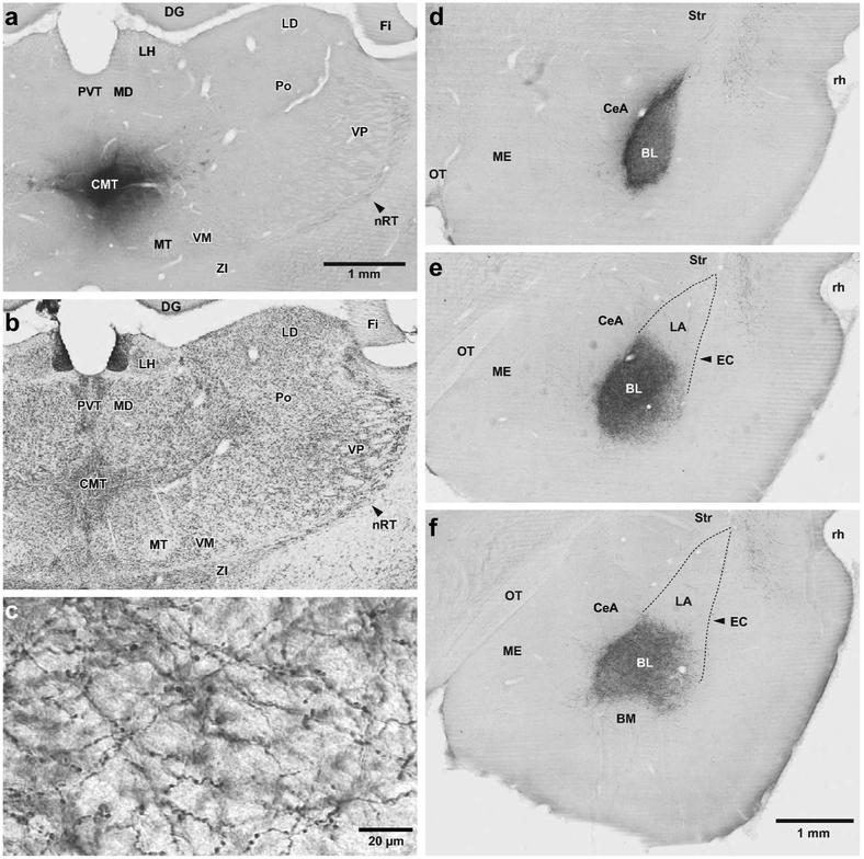
Figure 1.
CMT projections to the amygdala are largely confined to the basolateral nucleus (BL). (a,b) Two adjacent coronal sections processed to revealed the PHAL injection site in CMT (a) or the localization of thalamic nuclei in a Nissl-stained section (c) adjacent to that shown in a. (c) High power photomicrograph showing varicose PHAL-containing CMT axons ramifying in BL. (d-f) Three coronal sections showing the distribution of anterogradely labeled PHAL-immunoreactive CMT axons at rostral (d), intermediate (e), and caudal (f) levels of BL. Abbreviations: BL, basolateral nucleus of the amygdala; BM, basomedial nucleus of the amygdala; CeA, central nucleus of the amygdala CMT, central medial thalamic nucleus; DG, dentate gyrus; EC, external capsule; Fi, fimbria; LA, lateral nucleus of the amygdala; LD, laterodorsal thalamic nucleus; LH, Lateral habenula; MD, mediodorsal thalamic nucleus; ME, medial nucleus of the amygdala; MT, mammillothalamic tract; OT, Optic tract; nRT, reticular thalamic nucleus; Po, posterior thalamic nuclear group; PVT, paraventricular thalamic nucleus; rh, rhinal sulcus; Str, striatum; VM, ventromedial thalamic nucleus; VP, ventroposterior thalamic nucleus; ZI, zona incerta. Scale bar in a also applies to b. Scale bar in f also applies to d and e.

