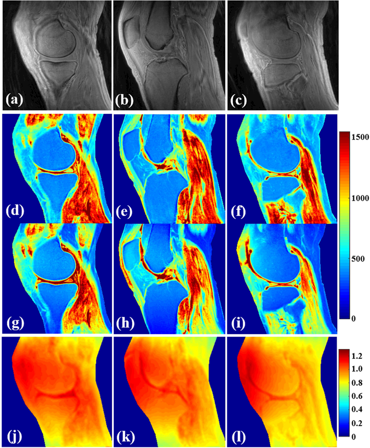
Figure 5.

Results in knee tissues from a healthy 35-year old male volunteer (a–l). (a–c) are the selected VFA images with FA = 5°. T1 mapping using both the proposed 3D UTE-Cones AFI-VFA (d–f) and B1-uncorrected VFA (g–i) methods are shown. The B1s maps generated by the AFI technique (j–l) are shown. B1 inhomogeneity induced T1 estimation errors in the images of g–i have been corrected by the proposed 3D UTE-Cones AFI-VFA method, especially in regions close to the coil boundary.
