
FIG. 6.
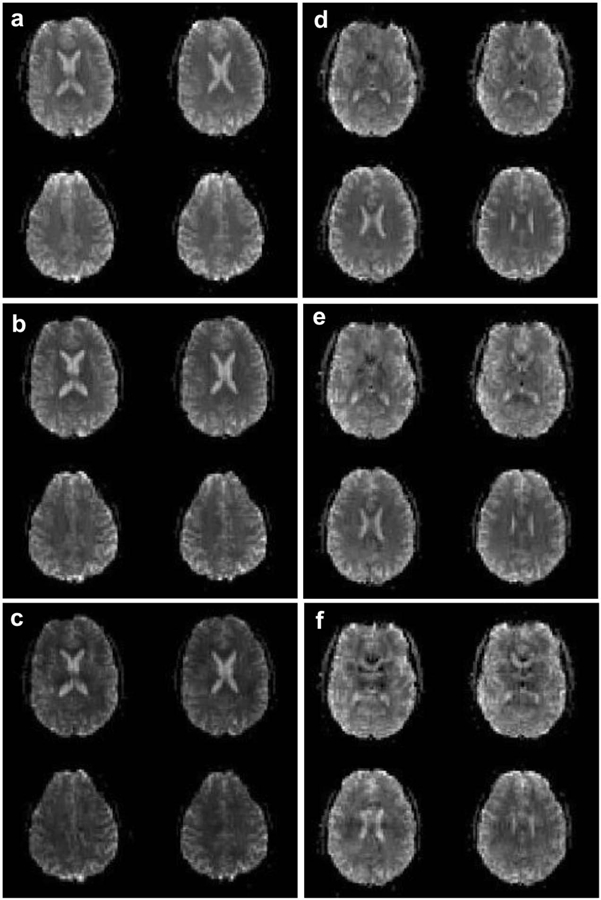
Four mean brain slices from an fMRI time series for a representative single subject acquired with (a) NSMS = 1, (b) NSMS = 4, and (c) NSMS = 8 SMS blipped-CAIPI CEPI. All three images are windowed identically. Also shown are the mean brain images for an (d) R = 1, (e) R = 4, and (f) R = 8 3D blipped-CAIPI CEPI fMRI time series. All three images are windowed identically.