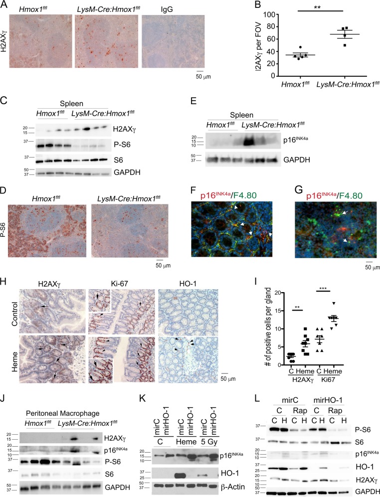
Fig. 2. Lack of HO-1 in myeloid cells leads to low phosphorylation of S6 and high expression of p16INK4a.
a, b Immunohistochemical staining with antibody against H2AXγ in the spleens from LysM-Cre:Hmox1fl/fl or Hmox1flfl mice. Quantification of the staining from n = 4–5 mice is shown in b. c Immunoblotting of the lysates of the spleens from LysM-Cre:Hmox1fl/fl or Hmox1flfl mice. n = 4 per group. d Immunohistochemical analysis of the phosphprylated S6 (P-S6) in the spleens from LysM-Cre:Hmox1fl/fl or Hmox1flfl mice. n = 4–5 per group. e Immunoblotting of the lysates of the spleens from LysM-Cre:Hmox1fl/fl or Hmox1flfl mice. n = 3 per group. f, g Immunostaining with antibodies against p16INK4a and F4.80, a marker of macrophages in the colonic (f) or spleen (g) tissues of C57/Bl6 mice. h, i Immunohistochemistry with antibodies against H2AXγ, Ki67 and HO-1 in the colon of mice treated with vehicle (control) or heme (35 mg/kg, i.p.) daily for 2 weeks. n = 4 mice per group. Representative sections are shown in h and quantification in i *p < 0.05, **p < 0.01, ***p < 0.001. j Peritoneal macrophages were isolated from LysM-Cre:Hmox1fl/fl or Hmox1flfl mice and lysated. Western blot was performed using n = 4 per group. k RAW 264.7 macrophages with stable knockdown of HO-1 were treated with heme (50 μM) or irradiation (5 Gy) for 6 h. Immunoblotting was performed using anti-HO-1 and anti-p16INK4a antibodies. Western blot is representative for n = 3 independent experiments. l RAW 264.7 macrophages with stable knockdown of HO-1 were treated with rapamycin (20 nM) for 15 min prior addition of heme (50 μM) for 6 h. Western blot is representative for n = 3 experiments

