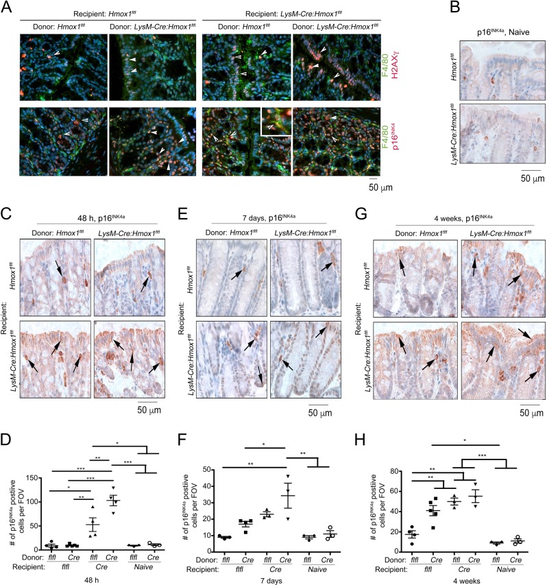
Fig. 4. Expression of p16INK4a in the colonic epithelium in response to genotoxic stress in HO-1 chimeric mice.
a Expression of p16INK4a (red) or H2AXγ (red) in Mφ (F4.80-green) in the colon after 10 Gy irradiation followed by BM Tx from LysM-Cre:Hmox1fl/fl or Hmox1flfl donor (D) to LysM-Cre:Hmox1fl/fl (LysM-Cre) or Hmox1flfl (flfl) recipient (R) mice at 48 h after BM-Tx as in Fig. 4. Naive- non-transplanted mice. Co-localization of p16INK4a and F4.80 is highlighted in inset (400x). b Immunohistochemical staining of p16INK4a in naive colons isolated from Hmox1flfl and LysM-Cre:Hmox1fl/fl mice. c–h Representative pictures of staining and quantifications of number of cells positive for p16INK4a in the colon after 10 Gy irradiation followed by BM Tx from LysM-Cre:Hmox1fl/fl (Cre) or Hmox1flfl (flfl) donor to LysM-Cre:Hmox1fl/fl (Cre) or Hmox1flfl (flfl) recipient (R) mice. 48 h (c, d), 7 days (e, f) or 4 weeks (g–m) after BM Tx colon tissues were harvested and stained. *p < 0.05; **p < 0.01, ***p < 0.001

