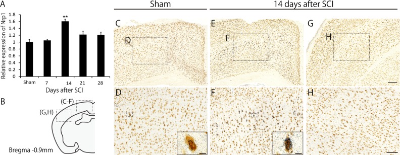
Fig. 2. Neuropilin-1 (Nrp1) is upregulated in the motor cortex in the pruning phase.
a RNA in the motor cortex was extracted at 1, 7, 14, 21, and 28 days after spinal cord injury (SCI) or sham treatment and was subjected to real-time PCR. Nrp1 expression at 14 days after SCI was significantly upregulated compared to that in sham mice. Data are presented as mean ± SEM. n = 3, **p < 0.01, one way ANOVA followed by Tukey-Kramer test. b Schematic diagrams of brain sections representing the approximate rostrocaudal level relative to bregma, −0.90 mm. c, e Representative images showing Nrp1 mRNA signals (blue) counterstained with NeuN (brown) in the motor cortex of sham (c) and injured (e) mice. In injured mice, more Nrp1 signals were observed. d, f Higher magnification images of the boxed regions in (c) and (e). The inset images in (d) and (f) are close-up view of the Nrp1 mRNA positive neurons marked in the boxed area. Scale bar: 10 μm. g, h In situ hybridization analysis for Nrp1 mRNA in the somatosensory cortex (g) and higher magnification of the boxed region in (g) in an injured mouse. Lower intensity of Nrp1 signals was observed in the somatosensory cortex compared to the motor cortex. Scale bar: 200 μm (g), 100 μm (h)

